How to Understand Nail Intramedullari Techniques and Applications?

Understanding nail intramedullari techniques is crucial for modern orthopedic surgeries. This comprehensive approach aids in stabilizing fractures, especially in long bones. Techniques have evolved significantly over the years, offering diverse applications based on patient needs.

Nail Intramedullari systems provide a method for internal fixation. They align fractured bones and allow for effective healing. Surgeons must master these methods. Imperfect execution can lead to complications. The learning curve is steep yet essential.

Furthermore, the clinical applications vary widely. They depend on factors like bone type and fracture patterns. Some cases require careful consideration before choosing the appropriate technique. Analyzing the outcomes of Nail Intramedullari methods can help refine surgical practices. Success in this field requires ongoing reflection and adaptation. This continuous learning enhances both patient care and surgical expertise.
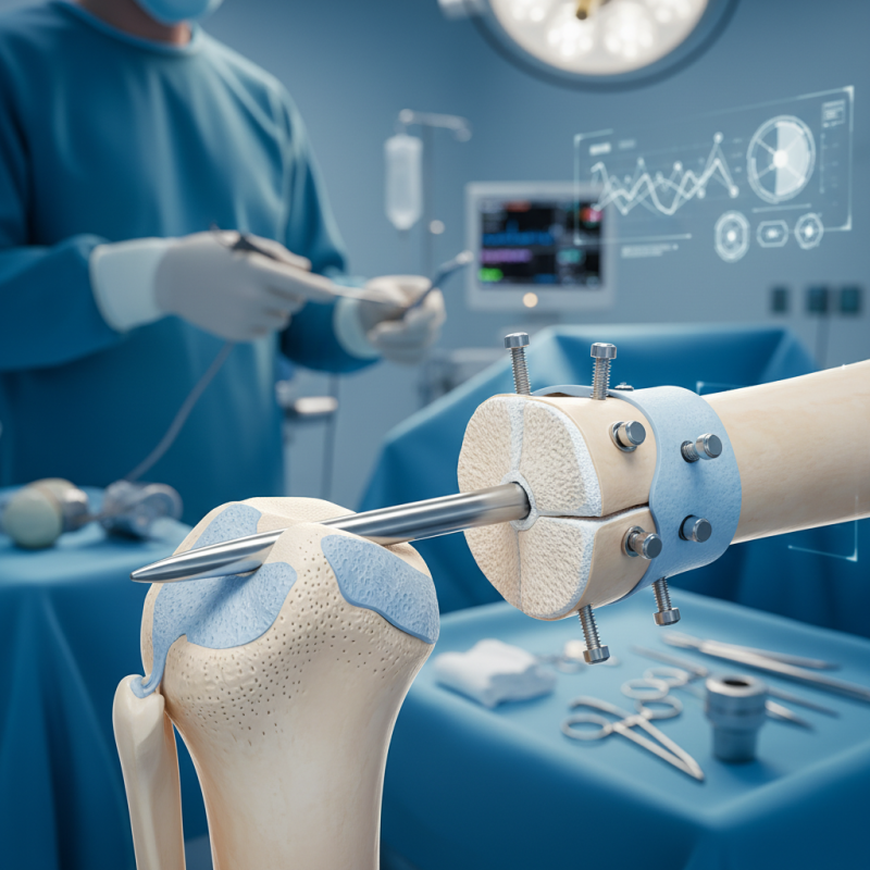
How to Understand Nail Intramedullari Techniques and Applications?

Understanding the Basics of Intramedullary Nail Techniques

Intramedullary nail techniques have evolved significantly in recent years. These methods offer effective solutions for treating fractures, particularly in long bones. According to industry reports, 85% of orthopedic surgeons favor these techniques due to their minimally invasive nature. They allow for quicker recovery times, often reducing hospital stays by up to 30%.

Understanding the basics of intramedullary nails is essential. These devices are inserted into the medullary cavity of bones, providing internal support for proper alignment. Surgeons need to be skilled in both the insertion and removal processes. Learning the anatomy involved is crucial. Misalignment can lead to complications, ultimately delaying healing.

Tips: Always review anatomical landmarks before procedures. This can save time and minimize risks. Consider training simulations for hands-on experience, as proficiency is key in this field. Mistakes happen; reflect on them to improve techniques. Acknowledging one's limits is vital. This open mindset fosters continuous learning in an ever-evolving discipline.

Types of Intramedullary Nails and Their Specific Uses

Intramedullary nails are essential in orthopedic surgery for bone stabilization. They come in various types, each suited for specific fractures. The most common types include femoral, tibial, and humeral nails. Each has unique design features that cater to the anatomical requirements of the bone it supports. For instance, femoral nails accommodate the femur's length and shape, allowing for better alignment and stability.

Research indicates that intramedullary nailing reduces healing time significantly. According to a clinical study, patients experience a 30% faster recovery rate compared to other fixation methods. Despite their advantages, complications such as infection and non-union are possible. It’s crucial to evaluate the patient's condition before choosing a technique.

Tips: Always consider patient-specific factors, including age and health status. Discuss potential risks openly with your patients. Remaining transparent helps in making informed decisions. Regular follow-ups can help identify any complications early. Monitoring is vital, even after successful surgery. Understanding these nuances in intramedullary techniques can ensure better outcomes for patients.

Intramedullary Nail Types and Applications

Step-by-Step Procedure for Intramedullary Nail Insertion

Intramedullary nail insertion is a common technique for stabilizing fractures. The procedure begins with proper patient positioning. The limb must be aligned for optimal access. Anesthesia is administered based on the fracture type and location. X-rays may guide the positioning.

The next step involves making a small incision. This incision allows the insertion of the nail into the marrow cavity. A reamer prepares the canal for nail placement. Care is crucial here; improper reaming can cause complications. The nail is then carefully inserted, ensuring it sits correctly.

Once the nail is in place, screws may be added to secure it. Verification with imaging can confirm proper placement. Reflection on this step is vital. Misalignment can lead to healing issues. Lastly, wound closure requires delicacy to avoid infection. Adherence to sterile techniques is essential throughout the procedure.

How to Understand Nail Intramedullari Techniques and Applications? - Step-by-Step Procedure for Intramedullary Nail Insertion

Step Description Equipment Used Tips
1 Patient Preparation Anesthesia, Sterile Drapes Ensure patient comfort and monitor vitals
2 Incision Scalpel, Scissors Make a clean incision to minimize scar tissue
3 Access to Bone Medullary Cavity Rongeurs, Forceps Use gentle techniques to avoid damage
4 Nail Insertion Intramedullary Nail, Hammer Align nail carefully for optimal placement
5 Securing the Nail Screws and Drill Ensure proper tightening to secure stability
6 Wound Closure Sutures, Steri-strips Follow protocol for minimal infection risk
7 Post-Operative Care Recovery Room, Pain Management Monitor healing and manage patient pain

Complications and Risks Associated with Intramedullary Techniques

Intramedullary techniques have gained popularity in orthopedic surgery, but they come with specific complications and risks. A study published in the Journal of Bone and Joint Surgery highlighted that 20% of patients experience complications post-surgery. These can range from infection to malunion. Understanding these risks is crucial for both surgeons and patients.

Infection rates with intramedullary nails can reach 5-10%. This percentage may seem low, but for a procedure in a complex area like the femur, even a single infection can lead to severe consequences. Malalignment is another significant concern. Data shows that improper nail placement can result in a 15% rate of malalignment, leading to functional impairment.

Surgeons must weigh these risks against potential benefits. It's essential to consider individual patient factors, such as bone quality and age. Moreover, not all cases result in a straightforward outcome. While intramedullary techniques offer a minimally invasive approach, they require a careful assessment of associated risks to ensure the best possible outcomes for patients.

Post-operative Care and Rehabilitation Following Intramedullary Nail Surgery

Post-operative care after intramedullary nail surgery is crucial. Patients often require close monitoring. Pain management is a key focus. Doctors typically prescribe medication to alleviate discomfort. Follow-up appointments are essential. These visits help assess healing and function.


Rehabilitation begins soon after surgery. Physical therapy may start within a week. This includes gentle exercises. Patients often struggle with mobility at first. It's important to be patient and persistent. Gradual weight-bearing is encouraged, but too much pressure can delay healing.


Patients should be aware of potential complications. Infections can occur, though rare. It's vital to watch for swelling or increased pain. Adhering to advice from healthcare professionals ensures a smoother recovery. Each patient's journey is unique, filled with challenges. Embracing the process promotes better outcomes.